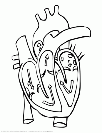

One of the most amazing organs in your body. Veins appear blue, but not necessarily because the blood in it is deoxygenated, rather because of the distortions of the vein itself and how we perceive the colors around it.
HEART BLOOD FLOW COLORING PAGE SKIN
So the dark red blood on the inside will be distorted, AND on top of this if we are looking at the vein surrounded by a reddish or tan environment such as the skin it will make it appear more blue (like if you were to put something purple next to something red, it would appear more blue).Īrteries are not red (if removed from the body and washed off) they are opaque tanish-white because of the wall of the artery itself is that color. Veins, on the other hand, are translucent, so what's inside will affect the coloring of what we see on the outside. Since arteries are opaque blood inside will not significantly affect its appearance in terms of coloring. In a healthy human, blood itself is always red, oxygenated blood will be bright red and deoxygenated blood will appear dark red.ĭon't forget that veins and arteries themselves have there own colors and consistencies, they are not completely transparent like glass, so what color you see on the outside isn't technically the same color as the inside fluid.Ī vein by itself has a translucent-whitish appearance if no blood is in it, and arteries are actually an opaque tanish-white color because of the thick layering of connective tissue on the outside called the adventitia. These two factors take away from the "bulge" argument.Īs of right now, I do not believe there is an answer to your question, but it definitely has to do with fetal development and early fetal circulation. My second problem is that the ductus arteriosis and the high resistance in the lungs limits the blood returning to the atrium via the pulmonary vein. I think the assumption is not grounded in any facts, but speculation. It seems that an assumed premise is both the tricuspid valve and mitral valve start out with three leaflets. I think there were two problems with this. This bulging forces two leaflets of the proliferating cell to join and form one leaflet The excess blood in the left atrium forces it to bulge to make room for excess blood Blood joins the left atrium from the regular sources (pulmonary veins) and also from the right atrium. During embryonic development pressures are higher in the right side of the heart than the left I only found one theory that makes some sense. I thought this was a really interesting question, and I did a little bit of research. The other valves are meant to be more easily opened so that blood can easily be pushed out of the heart. This means that there needs to be a reliable barrier between the Atrium and Ventricle which is why there are three flaps. So when the blood initially flows into the heart, it needs to be stopped in the Atrium and the Ventricle before being pushed out of the heart. The way these valves open is because the blood is literally being forced out of the Ventricle, and this is the main reason why they are different in structure. They have fibrous "strings" attached to the back so they can only open in one way. This is the same principle as the semi-lunar valves. However, you are not able to open the door in the opposite direction because the hinge will not allow the door to swing that way. With the turn of a knob, you are able to open that door and walk in.

To help envision this, imagine that you have a hinged door on the wall. These two valves are called this because they have two flaps of skin that can only open in one direction, again to prevent backflow. The other valves, the Pulmonary and Aortic valves are semi-lunar valves. Since they have three flaps of skin, they are called TRIcuspid (easy to remember). Imagine three flaps of tissue that come together to form a seal so that there is no back flow of blood. The Tricuspid valves (also called the atrioventricular valves) have a three flap design. This is a good question and in short the answer is no.


 0 kommentar(er)
